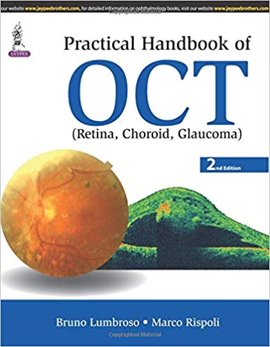
Optical coherence tomography (OCT) is a non-invasive imaging test that uses light waves to take cross-section pictures of the retina, the light-sensitive tissue lining the back of the eye (eyeSmart).
This new edition has been fully updated to provide clinicians and trainees with the most recent advances in OCT imaging. Beginning with an introduction to the technique for obtaining clear images, and discussion on normal anatomy, the following sections offer step by step guidance on the interpretation of OCT images and data acquired by OCT, ‘en face’ OCT and OCT angiography.
Each chapter has been revised and new examination and diagnostic protocols are covered in depth. The second edition includes two new chapters on OCT Dyeless Angiography and OCT Analysis and Interpretation of Common Disorders.
Highly illustrated with more than 400 clinical images and tables, this practical reference has been written by renowned experts Bruno Lumbroso and Marco Rispoli from Centro Oftalmologico Mediterraneo in Rome.
Key points
- Fully revised new edition providing latest advances in interpretation of OCT imaging
- Includes two new chapters and more than 400 clinical images and tables
- Authored by renowned ophthalmologists Bruno Lumbroso and Marco Rispoli
- Previous edition published in 2012

