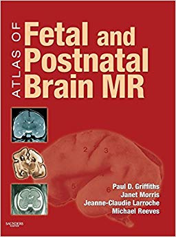
By Paul D. Griffiths FRCR PhD, Janet Morris MSc, Jeanne-Claudie Larroche MD
Hardcover: 272 pages
Publisher: Mosby; 1 edition (December 3, 2009)
Language: English
ISBN-10: 0323052967
ISBN-13: 978-0323052962
The Atlas of Fetal and Neonatal Brain MR is an excellent atlas that fills the gap in coverage on normal brain development. Dr. Paul Griffiths and his team present a highly visual approach to the neonatal and fetal periods of growth. With over 800 images, you’ll have multiple views of normal presentation in utero, post-mortem, and more. Whether you’re a new resident or a seasoned practitioner, this is an invaluable guide to the new and increased use of MRI in evaluating normal and abnormal fetal and neonatal brain development.
- Covers both fetal and neonatal periods to serve as the most comprehensive atlas on the topic.
- Features over 800 images for a focused visual approach to applying the latest imaging techniques in evaluating normal brain development.
- Presents multiple image views of normal presentation to include in utero and post-mortem images (from coronal, axial, and sagittal planes), gross pathology, and line drawings for each gestation.
Premium Content
Login to buy access to this content.What's Your Reaction?
Excited
0
Happy
0
In Love
0
Not Sure
0
Silly
0

