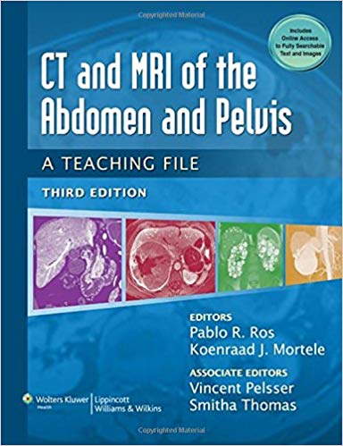
By Pablo R. Ros, Koenraad J. Mortele MD
Series: LWW Teaching File Series
Paperback: 204 pages
Publisher: LWW; Third edition (January 1, 2014)
Language: English
ISBN-10: 1451113528
ISBN-13: 978-1451113525
Now in its Third Edition, this trusted and practical volume in LWW’s Teaching File Series offers residents and practicing radiologists a unique opportunity to study alongside the experts in their field. For the first time, CT and MRI of the Abdomen and Pelvis is a hybrid publication, with a new paperback format and accompanying web content that includes a wealth of case studies users can access from their laptop, tablet, or mobile device. The book is useful both as a quick consult or study aid for anyone preparing for Board examinations in Radiology and other specialties where knowledge of CT and MRI of the abdomen and pelvis are required.
This skill-builder delivers…
- 413 structured case studies based on actual patients—each providing a brief patient history, as many as four CT/MR images, a short description of the findings, differential diagnosis, final diagnosis, and a discussion of the case.
- Detailed imaging of all areas of the abdomen and pelvis—including the liver and biliary system, pancreas, GI tract, spleen, mesentery/omentum/peritoneum, kidney and urinary system, retroperitoneum and adrenal glands, and abdominal wall—helps readers understand relevant anatomy and identify pathologies.
NEW to the Third Edition…
- Accompanying web site delivers access to the 150 cases from the print edition, plus 263 "bonus" cases for a total of 413 cases!
- 30% new cases address new challenges and provide timely information
Premium Content
Login to buy access to this content.What's Your Reaction?
Excited
0
Happy
0
In Love
0
Not Sure
0
Silly
0

