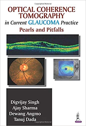
By Digvijay, M.D. Singh, Ajay Sharma, Dewang, M.D. Angmo
Paperback: 126 pages
Publisher: Jaypee Brothers Medical Pub; 1 edition (June 1, 2014)
Language: English
ISBN-10: 9789351521884
ISBN-13: 978-9351521884
ASIN: 9351521885
Glaucoma is a condition of the eye in which the optic nerve is damaged due to increased fluid pressure in the eye. Left untreated, the condition may lead to permanent blindness. Optical coherence tomography (OCT) is a non-invasive imaging test that uses light waves to take cross-section pictures of the retina, the light-sensitive tissue lining the back of the eye (geteyesmart.org). OCT is commonly used in the evaluation of patients with glaucoma. This manual is a concise guide to the use of OCT for the diagnosis of glaucoma. Beginning with an introduction to OCT, the book then provides in depth discussion on its use in glaucoma. Each of the following chapters describes the use of OCT for analysing associated parts of the eye, including the optic nerve, retinal nerve and ganglion cell, as well as macular and anterior segment OCT. The advantages and common pitfalls in OCT imaging and its interpretation are discussed at length. Key points Concise guide to OCT for diagnosis and evaluation of glaucoma Explains use of OCT for analysis of associated parts of the eye In depth discussion of advantages and common pitfalls in OCT imaging Includes more than 115 images and illustrations
Premium Content
Login to buy access to this content.What's Your Reaction?
Excited
0
Happy
0
In Love
0
Not Sure
0
Silly
0

