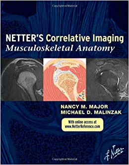
Musculoskeletal Anatomy is the first title in the brand new Netter’s Correlative Imaging series. Series editor and specialist in musculoskeletal imaging Dr. Nancy Major and coauthor, Michael Malinzak, presents Netter’s beautiful and instructive paintings and illustrated cross sections created in the Netter style side-by-side with high-quality patient MR images created with commonly used pulse sequences to help you visualize the anatomy section by section. With in-depth coverage and concise descriptive text for at-a-glance information and access to correlated images online, this atlas is a comprehensive reference that’s ideal for today’s busy imaging specialists.
- View upper and lower limbs in sagittal, coronal, and axial view MRs of commonly used pulse sequences, each slice complemented by a detailed illustration in the instructional and aesthetic Netter style.
- Find anatomical landmarks quickly and easily through comprehensive labeling and concise text highlighting key points related to the illustration and image pairings.
- Correlate patient data to idealized normal anatomy in the approximately 30 cross-sections per joint that illustrate the complexities of musculoskeletal anatomy.
- Scroll through the correlated images online at www.NetterReference.com.
Receive clarity and guidance for your practice, through the unique pairing of Netter art and clinical imaging

